Successful manoeuvres in the subarachnoid space
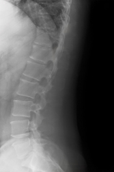
Spinal cord lesions are usually the result of trauma from injury but can be due to tumours, degenerative diseases or congenital conditions. Successful treatment often requires investigation of the affected area by insertion of a microendoscope into the area surrounding the spinal cord. The subarachnoid space is the cavity between between the arachnoid and the pia mater. The meninges consist of three layers: the dura mater, the arachnoid mater, and the pia mater. Members of the European project MINOSC made it their aim to develop a flexible microendoscope to visually assess the lesions and to operate simultaneously with associated equipment. In order to fulfil this objective, the physical, anatomical and physiological properties of the subarachnoid space must be analysed to ensure the accurate manoeuvrability of the endoscope. Project partners at Technion, the Israel Institute of Technology, specifically researched previously uncharted areas of the thoracic subarachnoid space. Magnetic resonance imaging (MRI) scans from 42 patients were merged to produce data on the dimensions of the spinal cord and the volume and shape of the dural sac and the subarachniod space. These were taken at mid- and inter-vertebral disc levels to produce a complete map. The measurements match those available from previous studies. They were recorded for the transversal plane which cuts the body into top and bottom portions. A low level of variance was evident as there was a high degree of symmetry. Another story was revealed in the sagittal plane however. Dividing the body into right and left portions, in the median plane, there was found to be a very significant coefficient of variation (as high as 42%). The findings in this study not only increase the knowledge of the structure of the spinal cord. The models also dictate the maximum dimensions of the endoscope so that it can perform accurate movements in the subarachnoid space. This will no doubt increase the success of surgery for those with spinal cord lesions.