Beam imaging rises to the challenge of sophisticated accelerators
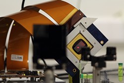
Beam diagnostics systems are essential elements of every particle accelerator. Without diagnostic elements, it would be impossible to operate linear accelerators for cancer radiotherapy, not to mention the world’s largest atom smasher, the Large Hadron Collider. They reveal the properties of a particle beam and how it behaves within the accelerator complex. The EU-funded project DITA-IIF(opens in new window) (Investigations into advanced beam instrumentation for the optimization of particle accelerators) was devoted to advancing the state-of-the-art of beam diagnostics based on light emitted by a beam of charged particles. ’There are many varieties of beam diagnostics, some based on direct detection of charges and currents induced by the beam. DITA-IIF was a research project whose aim was to advance the state-of-the-art of optical or near-optical radiation diagnostics,’ explained Professor Carsten Welsch, project coordinator from the University of Liverpool, United Kingdom. Optical radiation imaging The DITA-IIF team, led by Dr Ralph Fiorito from the Cockcroft Institute at the University of Liverpool, developed a new algorithm to analyse optical transition radiation produced when a beam intercepts a thin foil. Specifically, the shape of the measured image of optical transition radiation – the so-called point source function – from a single electron is used to calculate beam size. To achieve sub-micron accuracy, artificial artefacts such as aberrations and misalignments are isolated and removed. ‘The new method is currently used to improve the resolution of optical systems developed to image micron-sized beams that are produced by the Accelerator Test Facility at the High Energy Accelerator Research Organisation in Tsukuba, Japan,’ notes Professor Welsch. Furthermore, researchers have developed a new beam imaging technique relying on an array of electronically controlled micro-mirrors. This digital light processor offers an optical mask to filter out optical radiation from the core of the beam and add the resultant image to that of the outer halo. As a spatial filter, it mitigates light diffraction around the optics systems used to image the beam. To evaluate the performance of these beam imaging systems, researchers currently compare lab measurements with simulation results from the Zemax Optical Studio. Professor Welsch adds that ‘this comparative study will improve our understanding not only of the beam imaging method but also of any digital light processor used for medical or astronomical imaging as well as remote sensing applications.’ Non-invasive coherent radiation imaging The DITA-IIF team went a step further by proposing a new beam imaging method based on measurements of coherent radiation produced when a number of electrons pass through an aperture. Both the angular and spatial distribution of the coherent diffraction radiation is captured and analysed to retrieve the beam length. Importantly, researchers could image the point source function of coherent radiation diffraction in the terahertz wavelength regime. Using the 20 GeV electron beam generated at the Facility for Advanced Accelerator Experiment Tests (FACET) at Stanford University in the US, they also demonstrated the lack of any contamination from other radiation sources. Besides addressing challenges in high energy beam imaging, ‘the diagnostics developed within DITA-IIF have excellent prospects for applications to a wide variety of accelerators as well as light sources. They provide better resolution in the least invasive way and are a game-changer for fully characterising a charged particle beam,’ concludes Professor Welsch.


