New imaging technology could predict patient response to NMD treatment
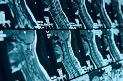
The project’s purpose was to enable the monitoring of patient response to novel therapies in neuromuscular diseases (NMDs), with DMD being used as a case in point. As Dr Andrew Blamire, Director of Newcastle University’s Centre for In Vivo Imaging, explains, there are two major driving factors for developing such monitoring technologies. The first is the need to identify statistically meaningful differences in response to treatment. ‘Many NMDs have a profound impact on muscle strength and function. In trials of new treatments, one will therefore assess, for example, the distance the patient can walk in 6 minutes as an indication of muscle damage and then see how this changes over time or with treatment,’ Dr Blamire explains. ‘On the other hand, imaging technology can quantify the degree of muscle damage directly and non-invasively, and provide valuable new outcome measures for trials.’ The second factor is the need to know how the drug interacts with the patient. To achieve this, the project team created imaging technologies which, via drug-labelling approaches, may in the future allow physicians to follow where the drug goes in the body. New scanning approaches When it came down to identifying an exemplar disease, the choice of DMD was a no-brainer. Consortium members shared a common interest in the disease and, at the time of establishing the BIOIMAGE-NMD programme, a number of new treatments for DMD were approaching clinical trial. The project team started their work with a comprehensive evaluation of several forms of magnetic resonance imaging (MRI). They developed an approach called diffusion imaging, along with new methods to analyse and quantify the scans. In parallel, they developed an approach using positron emission tomography (PET) – a form of imaging which uses radioactively labelled tracer molecules – to label a novel drug called antisense oligonucleotide (AON) which was about to be trialled in DMD patients. ‘We developed a way to add a radioactive atom to the drug and make it visible from outside the body using the PET scanner,’ Dr Blamire says. ‘We have shown in animal models that this new labelling approach does not prevent the drug from acting in the way in which it was designed. This is an important development, as AON are not just being used in patients with DMD but has a wider potential role in diseases with a genetic cause.’ BIOIMAGE-NMD’s new diffusion imaging advances have yet to be applied in clinical trials, however, the use of existing quantitative MRI scanning during AON clinical trials allowed for mapping DMD progression by imaging. ‘We observed that imaging can clearly detect subtle changes in disease progression and is more sensitive than the routine clinical assessment. We also observed that there is a lot of variation in the rate of disease progression between individual patients, even though they have very similar genetic changes causing their disease, and we identified that we need to further understand the cause of this variation to improve the utility of imaging as an outcome measure’. Though the project has now been completed, consortium partners are continuing to collaborate on the further evaluation and interpretation of the data collected during BIOIMAGE-NMD and are developing future plans for extension of the research. ‘All partners continue with separate (and collaborative) projects and are contributing imaging expertise into NMD trials being led by other commercial partners outside of the original BIOIMAGE-NMD consortium. We are considering all opportunities to obtain further funding for this research, but have yet to secure funding,’ Dr Blamire concludes.