An AI with hyperspectral vision can spot the early signs of retinal disease
More than 39 million Europeans are affected by retinal pathologies such as macular degeneration and diabetic eye disease(opens in new window). This number is expected to grow 25 % by 2050 due to ageing populations and rising levels of diabetes. Early diagnosis and treatment could prevent up to 98 % of sight loss resulting from diabetes.
Haemorrhages
The XpeFundus project was a market feasibility study for a new medical imaging technology that is sensitive to tiny changes in the biochemistry of retinal tissues. “The retina and layers beyond are full of veins, and diabetes affects veins, causing them to break and create haemorrhages,” explains project coordinator Vassilis Papadakis. “We are able to detect these haemorrhages and other tissue pathologies at an early stage.” Existing methods for diagnosing retinal pathology, such as optical coherence tomography (OCT), use a bright white laser light to illuminate the back of the eyeball, and the images generated require specialist training to analyse. This limits their use to hospital settings. In addition, OCT can only detect structures greater than 10 micrometres in depth.
Lost vision
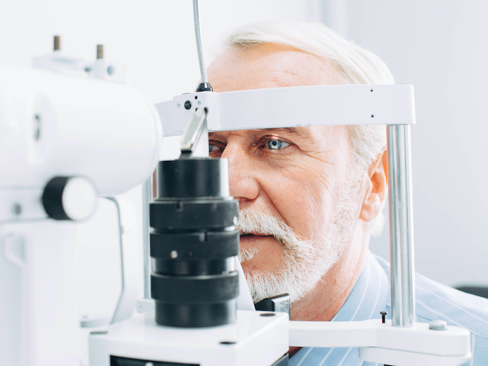
The imaging equipment developed by Papadakis and his team at Xpectraltek(opens in new window) illuminates the eye using progressive bands of light around 15-25 nanometres across. These layers are then interwoven and analysed by artificial intelligence (AI) to provide a diagnosis. The results is a biomolecular map where biochemical aberrations as small as 2 micrometres can be visualised. The equipment is designed for small clinics and optometrists, without the need for specialist training. By providing a test outside of a hospital setting, patients can be diagnosed sooner, says Papadakis. “A significant number of people don’t know they have retinal pathology, and by the time they visit a doctor they could have lost 20 % of their vision,” he says. “By then it might be too late to recover even a small percentage of that.”
Disruptive tech
XpeFundus was supported by the EU’s Horizon 2020 programme. “With start-ups there is always a problem of funding, if we were big we could do it ourselves but it’s impossible at this level,” notes Papadakis. “The EU grant gave us an opportunity to study the market better and develop our business plan, and to understand who our competitors were, who we should avoid and who we should reach out to.” Papadakis plans to apply for EC accelerator funding later this year to bring the device through clinical trials and onto the market. The hyperspectral imaging technology behind XpeFundus is patent protected and has previously been used to examine cultural artefacts(opens in new window) and to provide agricultural analysis(opens in new window) such as nutritional deficiencies, diseases and hydration levels in crops. “The idea came from my PhD 17 years ago, where I wanted to create something useful for the community,” adds Papadakis. “Together with my partners, I started Xpectraltek to achieve goals I could not through academia, to push disruptive and innovative ideas as products that can really be helpful to the community and people, into the market.”