Shedding light on infant brain health
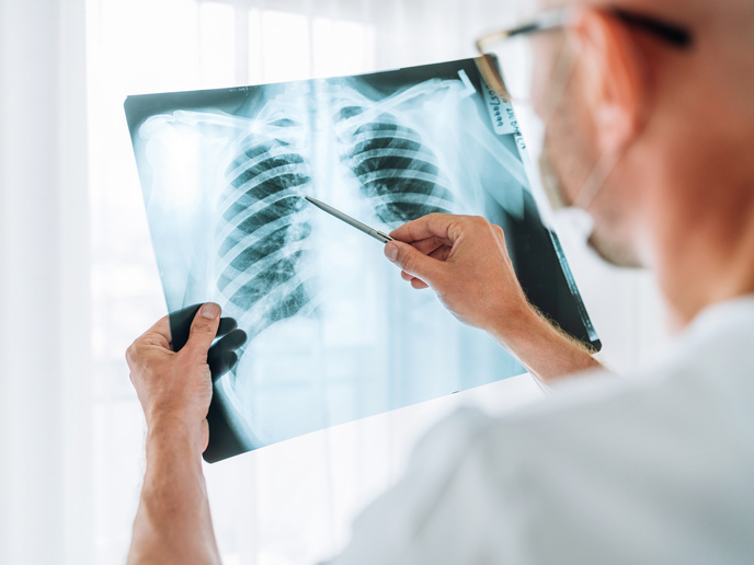
Infants born with a congenital heart defect(opens in new window) (CHD) often require complex surgeries within their first months of life. While these procedures are lifesaving, they carry a high risk of brain injury due to fluctuations in blood flow and oxygen supply. Traditional monitors provide only limited information, making it difficult for clinicians to understand how the brain responds during and after surgery.
Monitoring the infant brain
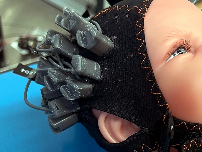
The EU-funded TinyBrains(opens in new window) project set out to change this by creating new tools that combine light-based and electrical measurements to study brain health in unprecedented detail. “Our goal was to develop non-invasive systems that allow real-time monitoring of brain function, oxygen consumption and blood flow in newborns with CHD,” explains project coordinator Turgut Durduran. “By doing so, we can provide clinicians with better data to guide life-saving decisions.” The TinyBrains team designed two innovative prototypes, a neuroimager and a neuromonitor, that merge three powerful technologies into one compact device: near-infrared spectroscopy (NIRS), diffuse correlation spectroscopy (DCS), and electroencephalography (EEG). NIRS and DCS use harmless near-infrared light to measure cerebral blood flow and oxygen metabolism, while EEG captures the brain’s electrical activity. These signals together reveal how neurons, blood vessels and oxygen, collectively known as the neurovascular unit, interact in the brain. The TinyBrains system uniquely provides co-localised, simultaneous measurements that quantify how well the brain is functioning and whether it receives enough oxygen to sustain that activity. Tailored headgear and hybrid sensors make the device suitable even for delicate neonatal use.
From the lab to the clinic
To validate the system, researchers first developed a preclinical model mimicking complex neonatal heart surgery using piglets, a well-established model for studying newborn brain function. This allowed them to simulate procedures such as cardiopulmonary bypass and deep hypothermic cardiac arrest, and to monitor how brain activity and oxygenation changed throughout the process. The platform showed high sensitivity to alterations in oxygen delivery and neuronal response. TinyBrains then tested its devices in 29 infants undergoing cardiac surgery. The neuromonitor successfully captured how brain oxygenation and electrical activity evolved during different surgical phases, from the onset of cardiopulmonary bypass to recovery. Meanwhile, the neuroimager was used to study how babies’ brains responded to simple sounds, offering new insights into neurovascular coupling and developmental function before and after surgery. “These results demonstrated that our platform could track subtle changes in brain function that other clinical monitors miss. This is a critical step toward preventing neurological complications in these vulnerable infants,” highlights Durduran.
Transforming neonatal care
The project’s greatest achievement lies in creating a comprehensive, non-invasive and multimodal tool for studying the infant brain. For the first time, clinicians can simultaneously assess neuronal activity, oxygen consumption and blood flow, offering a complete picture of cerebral health, known as the ‘brain health index’. The seamless data integration pipelines of TinyBrains make the technology compatible with hospital environments, facilitating adoption by clinical teams. The team is now working on a clinical roadmap to bring these prototypes closer to real-world use. This includes multi-site clinical trials, collaborations with technology developers and continued research into developmental neuroscience.