Seeing deeper into the human brain
Understanding the human brain in health and disease remains one of the great scientific challenges of the twenty-first century, with profound medical, societal and economic implications. Magnetic resonance imaging (MRI) has become a cornerstone of this effort because it can map brain anatomy, function and connectivity non-invasively. However, to detect finer details, researchers need stronger magnetic fields and more sensitive scanners than the 1.5 tesla systems currently operating in most hospitals. This ambition led to the creation of the Iseult scanner(opens in new window), an ultra-high-field MRI system developed in France to operate at 11.7 tesla and investigate the human brain at unprecedented scale.
Launching the investigations
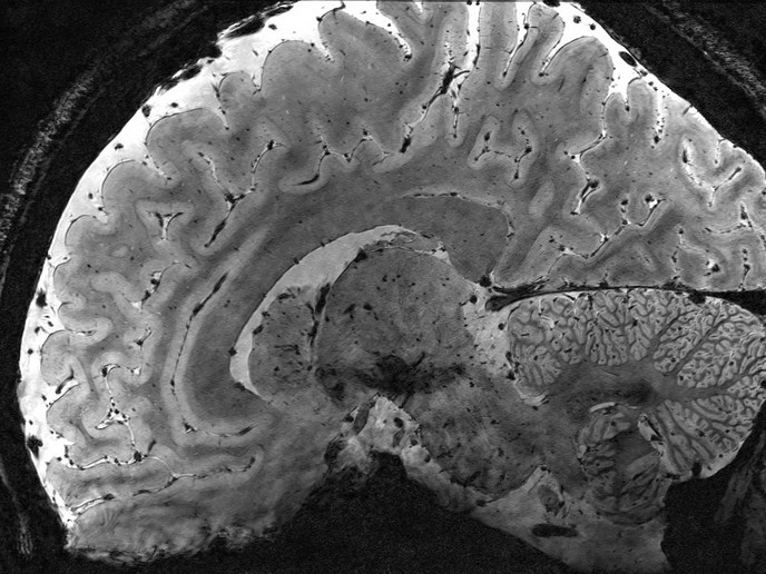
The EU-funded AROMA(opens in new window) project brought together six partners from five countries across academia and industry to help launch human brain studies on the Iseult 11.7 tesla MRI scanner. Higher magnetic field strengths generate stronger signals, which can be exchanged for finer spatial or temporal resolution. “The 11.7 tesla MRI scanner is designed to act as a non-invasive microscope, opening a unique window onto brain structure and function,” states project coordinator Nicolas Boulant. Researchers were able to acquire anatomical images at dimensions reaching the mesoscale of brain organisation(opens in new window), allowing visualisation of smaller anatomical features. The gains are not only about detail, but also speed. A scan lasting just over 4 minutes at 11.7 tesla would require dramatically longer acquisition times at lower field strengths to achieve comparable image quality.
Overcoming engineering challenges
Operating at such extreme field strength created substantial technical obstacles. Radio frequency coils had to efficiently excite water molecules and receive signals to leverage the scanner’s potential. Mechanical vibrations and interactions between components also threatened image quality. Motion during scanning posed another problem, especially at ultra-high resolution. To address this, the AROMA consortium developed advanced motion-correction methods(opens in new window) that significantly improved success rates. To validate the feasibility of human imaging at 11.7 tesla, 20 volunteers were scanned, with no adverse effects reported.
New paths in neuroscience
With these tools in place, the scanner is beginning to support functional MRI studies, including experiments resolving activity across the depth of the cerebral cortex. This could help researchers understand how information is processed across different cortical layers. “The generated images have been inspiring to the neuroscience community and show that the mesoscale world of the human brain is at our fingertips,” highlights Boulant. Potential applications also include neurodegenerative and psychiatric disorders such as Alzheimer’s disease, Parkinson’s disease, multiple sclerosis, schizophrenia and depression. Researchers hope the extra sensitivity may reveal subtle biological changes that remain invisible at lower field strengths. The platform is intended to be open to external users, although further work is still needed to expand the portfolio of MRI sequences that can be safely and efficiently deployed. “AROMA was a pioneer project that overcame many barriers of scanning at 11.7 tesla. The next challenge is to convert this technical success into clinically useful discoveries,” concludes Boulant.