Bioprinting at the speed of light
Drug development is a laborious and costly process requiring billions of euros and at least 10 years on average to develop one clinically applicable drug. Therefore, it is pivotal to improve the predictive power of preclinical screening to remove ineffective candidates as early as possible using 3D in vitro models that summarise the extent of native organ complexity and function. Bioprinting technologies are transforming how researchers design human tissue models for regenerative medicine and pharmaceutical research. However, current methods have their limitations as they follow a slow layer-by-layer process. As a result, building tissues larger than a cubic centimetre can take hours, far too long for sensitive cells to survive and function properly.
Bioprinting with light
To address the limitations of existing bioprinting techniques, the EU-funded ENLIGHT(opens in new window) project introduced a new approach called volumetric bioprinting. Inspired by medical tomography, this method utilises light patterns to shape cell-containing biomaterials into living functional tissues. By placing the biomaterial on a rotating platform and projecting a different pattern of light with every angle of rotation, functional living tissues are produced. The combination of the patterns from each angle ensure that only specific regions solidify, resulting in a specific 3D design. Because this process uses visible light and no physical pressure, it avoids damaging the cells and supports high survival and function rates. “We can print centimetre-sized tissues in just 10 seconds, using a light-based process that is exceptionally gentle on cells,” explains project coordinator Riccardo Levato.
A new ‘mini-pancreas’ for diabetes drug screening
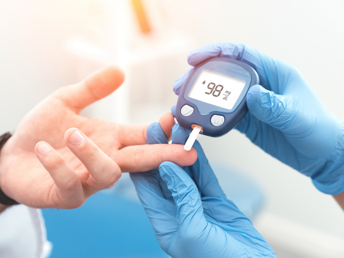
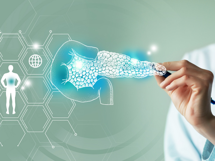
One of the project’s most promising applications is a bioprinted pancreas model. The team used a gelatin-based hydrogel, derived from collagen and encapsulated pancreatic islets, generated from patient-derived induced pluripotent stem cells. These islets are rich in insulin-secreting beta cells, which are lost or malfunction in people with Type 1 diabetes (T1D). The 3D model was designed with porous architecture to mimic natural blood vessel flow. Placed in a perfusion bioreactor, the model allows researchers to simulate blood circulation and test how the islets respond to drugs or toxic compounds. Importantly, because the cells are patient-specific, the system can be employed for personalised medicine. “We have screened different anti-diabetic drugs, as well as molecules that could cause toxicity in the pancreas. The goal is to make drug testing more efficient and effective, as we build a reliable and reproducible system that is based on human cells,” highlights Levato.
Impact and future directions
ENLIGHT demonstrated for the first time that volumetric bioprinting can enhance the function of complex tissues, like pancreatic islets, for advanced research applications. The bioprinter has been launched on the market by partner Readily3D(opens in new window), while a novel set of gelatin-derived materials for 3D printing and tissue culture have been commercialised by Rousselot(opens in new window), also a project partner. The team will continue to refine their printed models and explore their use for gene therapy testing, another area where current models fall short. They are also assessing how the technology can support cell transplantation therapies for T1D, using large-scale printed tissues as replacements for failing pancreatic islets. As bioprinting evolves, ENLIGHT’s technology could become a cornerstone for both drug development and regenerative medicine. It could help pharmaceutical companies identify safe and effective drugs earlier, reducing costs and the need for animal experimentation.