A tumour twin to track metastasis in real time
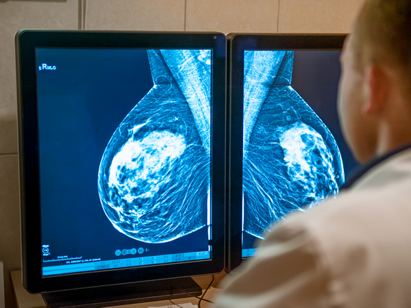
Metastasis to lymph nodes is often an early and decisive step in cancer progression. However, the complex dialogue between primary tumours and lymph nodes remains poorly understood, limiting the development of effective diagnostic tools and anti-metastatic therapies. Addressing this gap is essential, particularly for lung cancer patients, where lymph node involvement strongly influences prognosis and treatment decisions.
A biological twin of the patient
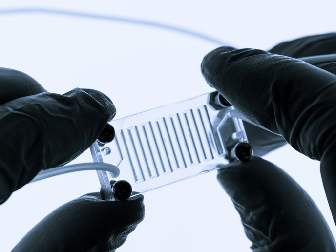
The EU-funded Tumor-LN-Oc(opens in new window) project set out to recreate the critical interaction between the primary tumour and the lymph nodes in a controlled, patient-specific setting. The consortium developed a tumour-lymph node-on-chip platform to monitor metastatic processes in real time, as well as identify molecular and spectral signatures of migrating cells. The same system can be used for parallel drug testing. The platform contains surgically removed primary tumour slices and matched lymph node cells from the same lung cancer patient. These are placed in separate compartments on a microfluidic chip, which communicate through microchannels. Artificial cilia(opens in new window) generate continuous recirculatory flow and a controlled gradient, recreating physiological conditions. “The tumour-lymph node-on-chip is designed to act as a biological twin, recreating the environment where cancer cell migration often begins,” explains project coordinator Ioanna Zergioti.
Capturing cancer cell migration in real time
A key innovation of Tumor-LN-oC lies in its integrated imaging and analytical modules. The micro-optics system produces images of micrometre optical resolution. It monitors whether tumour cells start moving, how fast they move, and their direction of movement. Machine learning algorithms quantify motion, providing real-time data on speed and directionality. In parallel, an infrared spectroscopy module enables molecular characterisation, generating spectral fingerprints of migrating or metastasising cells in the chip’s channels. As a result, the platform has the potential to identify diagnostic signatures that could later be applied to tumour and lymph node biopsies. “Biologically, one clear finding has emerged: lymph node-derived signals promote tumour cell migration,” highlights Zergioti. The consortium observed that tumour cells can move towards stronger signals released by lymph node cells, and that this directional guidance can be disrupted when the sensing mechanism is blocked. Such insights could inform strategies to inhibit metastatic spread.
Clinical validation
The Tumor-LN-oC platform has been clinically validated using samples from 60 lung cancer patients. Primary tumour slices and paired lymph node cell suspensions were co-cultured on the chip for several days, and soluble factors were analysed using sensitive proteomic and molecular approaches. Ongoing analysis is expected to identify biomarkers associated with metastatic potential. Importantly, patterns of tumor–lymph node interaction and migration in the chip were influenced by clinical features such as tumour stage, suggesting relevance to patient stratification. The consortium is also exploring the platform for preclinical drug testing, exposing patient-derived tissues to different therapeutic agents. This approach will help assess which treatments can suppress invasive behaviour.
Future impact
Tumor-LN-oC has turned the conceptual idea of personalised metastasis modelling into a tangible platform. Next steps include making the system more robust for clinical integration. Looking ahead, the platform could serve as a decision-support tool alongside standard diagnostics. If validated in larger studies, it may help oncologists stratify patients by metastatic risk and prioritise therapies most likely to prevent disease progression.